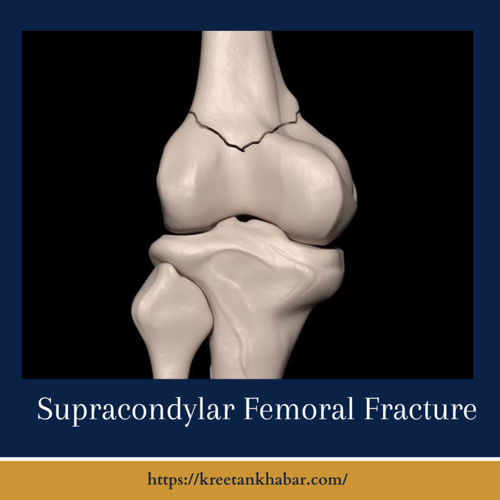
Supracondylar Femoral Fractures
Introduction
In the intricate world of orthopedics, the supracondylar femoral fracture emerges as a challenging terrain, demanding a nuanced understanding of the thigh’s complex architecture. This type of fracture, occurring just above the knee joint, brings forth a cascade of considerations, from its origins and classifications to the intricacies of diagnosis and treatment. Let’s embark on a journey through the orthopedic landscape to unravel the nuances of supracondylar femoral fractures, exploring the anatomy, causes, and the strategies employed by orthopedic specialists to guide patients on the path to recovery.

The Anatomy Unveiled
At the intersection of strength and mobility lies the femur, the body’s largest and strongest bone. The supracondylar region, located just above the knee joint, plays a pivotal role in weight-bearing and intricate movements. When this region bears the brunt of excessive force or trauma, the result can be a supracondylar femoral fracture, disrupting the harmonious function of the lower extremity.
The anatomy of a supracondylar femoral fracture reveals itself as a complex interplay within the lower extremity’s architecture. Imagine the femur, the body’s largest and strongest bone, as the central character, with the supracondylar region just above the knee joint serving a pivotal role in weight-bearing and movement. In the narrative of a supracondylar femoral fracture, this region becomes a crucial setting where forces, both external and internal, play out.
Trauma acts as the catalyst, disrupting the delicate balance and integrity of this intricate structure. The fracture, often occurring just above the knee joint, introduces a plot twist, challenging the stability and function of the lower limb. To truly understand the anatomy unveiled in a supracondylar femoral fracture, one must delve into the depths of this orthopedic narrative, where each element contributes to the intricate dance of bone, muscle, and joint dynamics.
Origins and Causes
Supracondylar femoral fractures unfold as a narrative influenced by various forces and factors, each contributing a unique chapter to the orthopedic story. Picture trauma as the catalyst, a forceful event such as a fall, sports-related injury, or an automobile accident setting the stage for the fracture’s emergence. Within this narrative, osteoporosis subtly becomes an underlying theme.
weakening bone density and adding a layer of vulnerability, especially in the aging chapters of life. Age itself becomes a factor, influencing the fracture narrative, as the resilience of the femur diminishes with the passage of time. In this intricate dance of origins and causes, the supracondylar femoral fracture story is written, weaving together the threads of force, bone health, and the passage of time in the orthopedic tapestry.
- Trauma as the Catalyst:
- Supracondylar femoral fractures often find their origins in traumatic events, frequently occurring due to high-impact forces such as falls, automobile accidents, or sports-related injuries.
- The fracture may involve varying degrees of severity, from hairline cracks to more complex fractures with displacement.
- Osteoporosis as an Underlying Theme:
- In some instances, underlying conditions like osteoporosis, characterized by weakened bone density, may contribute to the susceptibility of supracondylar femoral fractures.
- Weakened bones are more prone to fracture under stress, even with forces that may be considered moderate in healthy bone.
- Age as a Factor in the Fracture Narrative:
- Age becomes a key factor in the fracture narrative, with elderly individuals more susceptible to supracondylar femoral fractures.
- The bone’s density and resilience diminish with age, amplifying the risk of fractures, especially in the presence of pre-existing conditions.
Diagnosis: Unraveling the Fracture Story
Diagnosing a supracondylar femoral fracture is akin to unraveling a compelling mystery within the orthopedic realm. The narrative begins with a thorough clinical examination, where orthopedic specialists meticulously explore the patient’s medical history and scrutinize the details surrounding the incident. This preliminary chapter sets the stage for the visual narrative, portrayed through imaging studies like X-rays and CT scans.
These images unfold the fracture’s location, intricacies, and potential displacement, providing a comprehensive roadmap for orthopedic interventions. Much like skilled detectives, orthopedic professionals use these diagnostic tools to piece together the fracture story, ensuring that the subsequent chapters of treatment are tailored with precision to guide the patient on their journey toward recovery.
- Clinical Evaluations as the Preliminary Chapters:
- Diagnosis embarks on a journey of clinical evaluations, where orthopedic specialists delve into the patient’s medical history, scrutinize the details of the incident, and perform physical examinations.
- This initial phase sets the groundwork for a more comprehensive understanding of the fracture.
- Imaging as the Visual Narrative:
- The diagnostic narrative unfolds with imaging studies, such as X-rays and CT scans, providing a visual narrative of the fracture’s location, extent, and potential displacement.
- These imaging techniques offer a detailed roadmap for orthopedic interventions.
Treatment: Crafting the Road to Recovery
Addressing the intricacies of a supracondylar femoral fracture demands a personalized treatment narrative woven with precision and care. Picture the early chapters where conservative measures take the lead, delicately immobilizing the fracture with a cast or brace. This phase sets the stage for the orthopedic turning point, where surgical interventions become the protagonist for more complex fractures, orchestrating a symphony of realignment and internal fixation.
As the story progresses, the rehabilitation epilogue emerges, casting physical therapy as the hero guiding individuals through exercises to regain strength and flexibility. This comprehensive treatment narrative not only mends the fracture but also endeavors to restore the intricate choreography of the lower extremity, allowing patients to step back into the rhythm of their daily lives with renewed strength and mobility.
- Conservative Measures in the Early Chapters:
- The early chapters of treatment often involve conservative measures, especially for less severe fractures.
- This may include immobilization with a cast or brace, coupled with pain management and physical therapy to promote healing and restore mobility.
- Orthopedic Intervention as the Turning Point:
- For more complex fractures or cases involving significant displacement, orthopedic intervention takes center stage.
- Surgical procedures, such as open reduction and internal fixation (ORIF), aim to realign the fractured fragments and secure them in place with hardware.
- Rehabilitation as the Epilogue:
- The epilogue of treatment unfolds in the rehabilitation phase, where patients engage in physical therapy to regain strength, flexibility, and functional mobility.
- Rehabilitation is crucial for preventing complications and facilitating a smooth return to daily activities.
Conclusion: The Orthopedic Narrative Continues
Supracondylar femoral fractures carve a distinct chapter in the orthopedic narrative, emphasizing the delicate interplay between trauma, bone health, and age. Through advancements in diagnostic techniques and orthopedic interventions, the story of recovery is continuously evolving, with each patient’s journey adding depth to our understanding of the orthopedic landscape. As we navigate this terrain, the goal remains steadfast—to guide individuals towards healing, restoring the symphony of movement, and allowing them to resume their roles in the narrative of daily life.
Read also : Exploring the Delightful Boost of the Green Tea Shot 2023